


A few weeks ago, we wrote about a quiet flaw sitting in the middle of every DNA-encoded library (DEL) screen: a PCR step that doesn't treat every barcode fairly, and a bias problem that can make a real hit look identical to a well that just didn't amplify. We closed that piece with a promise: DEL isn't the only pooled, PCR-amplified selection method with this vulnerability. The same dynamic shows up in RNA display, and we'd dig into it separately.
RNA display (mRNA display, ribosome display, cDNA display, and their variants) is one of the most productive ways for drug discovery teams to find peptide and protein binders against targets that small molecules and antibodies can't touch. It's cell-free, it's fast, and it can search through libraries of trillions of variants in a single tube. It's also built on the same iterative loop, over and over: select, amplify, select again. Buried inside every one of those loops is a PCR step that has to be re-tuned by hand at, every single round, or the whole campaign quietly drifts off course.
This post is a primer on RNA display, where PCR sits inside it, and why a problem the field openly documents, but has mostly learned to work around by hand, deserves a different kind of fix.
What is RNA display, exactly?
RNA display is a family of cell-free selection methods that link a molecule's "genotype" (its encoding genetic sequence) directly to its "phenotype" (the peptide or protein it produces), so that a single physical entity carries both: no cells, no colonies, no phage required.
The most common variant, mRNA display, starts with a DNA library encoding trillions of peptides or protein variants. That library is transcribed into mRNA, each mRNA molecule is chemically tagged with puromycin, and in vitro translation covalently fuses the resulting peptide to the very mRNA that encoded it (Wikipedia, mRNA display; PMC8725378). Two close siblings adapt the same idea: cDNA display reverse-transcribes the mRNA into a more stable cDNA-protein conjugate before selection, and ribosome display skips puromycin entirely, keeping the nascent protein, its mRNA, and the ribosome itself stalled together as one intact complex during selection (Plückthun lab).
Whichever variant, the screening logic is the same: the genotype-linked library is incubated against an immobilized target, unbound material is washed away, and whatever survives is presumed to be enriched for binders. Campaigns commonly screen up to 10¹³–10¹⁵ unique variants, run 6–10 iterative rounds of selection with increasing stringency, and can deliver binders in the nanomolar-to-picomolar range; this is the core technology behind platforms like PeptiDream's PDPS and the RaPID system, used industry-wide against targets considered "undruggable" by conventional modalities (IRBM; PeptiFinder).
Where does PCR fit into an RNA display workflow?
After each round of selection, the surviving genotype-linked molecules are present in vanishingly small quantities. Before that information can move to the next round (or, after the final round, to sequencing), it has to become amplifiable DNA again. For mRNA and ribosome display, that means a reverse-transcription step first, since the recovered molecule is RNA, not DNA; cDNA display already did this before selection. Either way, the workflow converges on the same hinge point: reverse-transcribe if needed, then PCR-amplify the surviving pool, before feeding it into the next round of selection or into next-generation sequencing (NGS) to call which sequences were enriched (PMC8725378).
Here's what makes RNA display different from a single-shot selection method: that PCR step doesn't happen once. It happens at the end of every round, typically six to ten times over the course of one campaign, with the output of each round's PCR becoming the input to the next round's selection.
Why is PCR such a problem for RNA display specifically?
Every PCR reaction amplifies templates unevenly. RNA display has a few characteristics that turn that ordinary bias into something closer to a structural flaw:
The PCR step repeats every round, not once. A typical campaign runs 3–10 iterative selection rounds, each ending in its own amplification step before the next round begins (PMC8725378; IRBM). Bias introduced in round one doesn't stay in round one; it becomes part of the input distribution for round two, and round three, compounding with each pass.
Cycle count has to be re-guessed every round, by hand. Published mRNA display protocols instruct researchers to run a pilot PCR at several arbitrary cycle counts, check a gel, and visually pick the number of cycles where the band "just reaches a plateau, yet side products are minimized" (PMC5993046). Ribosome display protocols spell out that this number has to shift as the campaign progresses: 32–40 cycles in round one, dropping to around 25 by later rounds, entirely by hand, entirely by prior experience (University of Zurich protocol). Get the retitration wrong in any single round, and that round's error becomes the next round's starting point.
You can't just re-run a selection round to get more material. Whatever survived that round's wash is what you have to work with at the PCR step; there's no going back to collect more.
Parallel conditions guarantee unequal starting material every round. Labs routinely run multiple targets, mutants, or off-target counter-screens side by side in the same 96-well plate (Max Planck 96-well selection protocol), meaning every well carries a different amount of recoverable material into a PCR step that's usually run under one fixed program for the whole plate.
The field has already named the failure mode directly. Independent groups have documented "highly-expressed parasite-like" sequences that preferentially amplify in mRNA-displayed libraries regardless of their true starting abundance, first reported over twenty years ago and still actively studied today (Nature Scientific Reports). One doctoral thesis working directly with mRNA display selections found a nucleotide bias in the library that "may have been non-specifically enriched round-by-round as a result of inherent PCR amplification bias" (White Rose eTheses). That's language that should sound familiar to anyone who read our DEL post.
Overcycling and undercycling: a problem that compounds every round
The same exponential-amplification math that makes PCR powerful also makes it dangerous at both ends of the cycle count. In RNA display, that danger doesn't just happen once per campaign—it happens up to ten times.
Undercycle a low-abundance round, and true binders present at low copy number never reach detectable levels. They read as absent, and because the surviving pool becomes the next round's starting library, they don't just disappear from that round's data. They're gone from every round after it, too.
Overcycle a round to rescue low-abundance wells, and the higher-abundance sequences on the same plate blow past exponential phase into plateau, where PCR duplicates, chimeric products, and "parasite" sequences accumulate disproportionately. One methods paper puts it plainly: a critical failure mode in these workflows is "over-amplification that could result in preferential enrichment of some sequences before the selection [is complete]" (PMC4263282), meaning a mistuned PCR step can manufacture a false winner before the biology of the next selection round ever gets a vote.
There is no fixed cycle number that avoids both failure modes on a plate carrying multiple selection conditions with different recoverable yields; that same guess then has to be made again next round, as yield shifts with every pass of enrichment.
What would fixing this actually require?
Structurally, the fix has to happen at the amplification step itself, not downstream in the bioinformatics. Deduplication and chimera-filtering pipelines can clean up some artifacts after sequencing, but they can't recover a binder that dropped below detection during PCR in round three, and they can't undo the distortion that a mistimed cycle count already baked into round four's starting material.
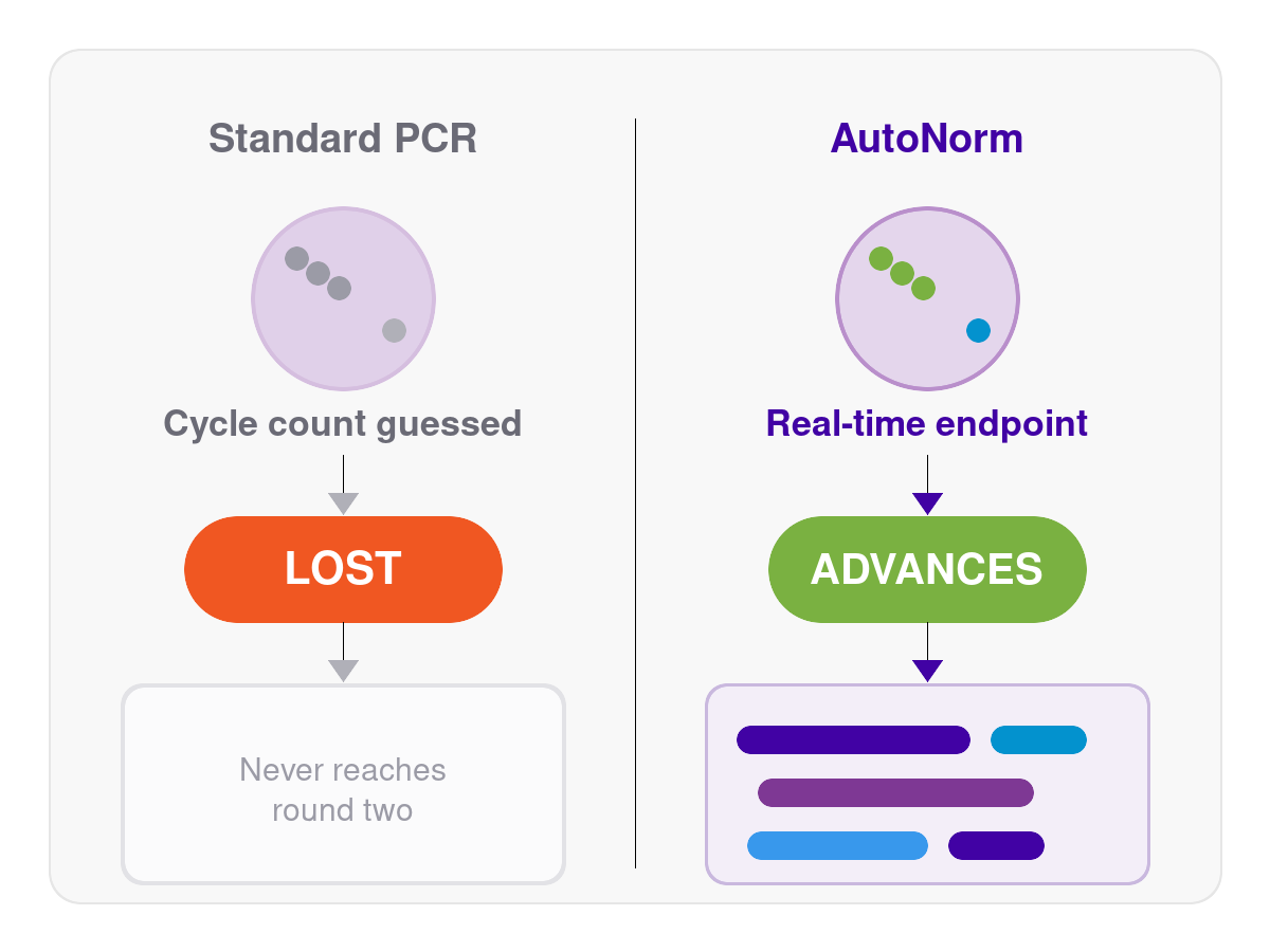
What the problem actually calls for is amplification that responds to each well's own starting material in real time, every round: stopping low-abundance wells once they've genuinely reached a usable signal, and stopping high-abundance wells before they drift into plateau-phase artifacts, without a person presetting one cycle number for the whole plate and then re-guessing it next round.
That's the exact mechanism iconPCR™ with AutoNorm was built to provide. iconPCR gives every well on a plate independent temperature control and real-time fluorescence monitoring, so each well stops individually the moment it reaches its own optimal endpoint, instead of every well on the plate sharing one fixed cycle count regardless of how much material it started with (n6 Products). On icon96™, that's 96 independently monitored wells per plate: enough to carry a full set of parallel selection conditions through a round side by side, with each one amplified on its own terms rather than forced through the same pilot-gel guess as its plate-mates.
This is exactly the class of problem we built iconPCR to solve, and it's one we've already started to put to work on the DEL side of this same selection-and-amplify category. RNA display's structure (the same PCR-and-hit-calling logic as DEL, but repeated six to ten times with a fresh cycle-count guess required every round) makes it, if anything, an even more natural fit for a smart thermocycler that finds its own endpoint instead of waiting to be told one.
The takeaway
RNA display is a genuinely powerful way to find binders against targets nothing else can touch, and PCR is the step quietly determining how much of a selection round's true signal survives to the next one. A binder that doesn't amplify cleanly isn't a failed molecule: it's a failed cycle count, and in an iterative selection method, that failure doesn't stay contained to one round. Until the amplification step can respond to what's actually happening in each well, some of the best binders in your library may never make it to round two, let alone to a final hit list.
If you're running mRNA, ribosome, or cDNA display selections and want to talk through where adaptive, per-well amplification could fit into your workflow, reach out to n6. We'd love to compare notes.
FAQ
What is RNA display?
RNA display is a family of cell-free selection methods, including mRNA display, ribosome display, and cDNA display, that physically link a molecule's genetic sequence to the peptide or protein it produces, allowing trillions of variants to be pooled, screened against a target, and identified later by sequencing rather than by testing one variant at a time.
How does RNA display screening work?
A genotype-linked library is incubated against an immobilized target, unbound material is washed away, and the surviving molecules are reverse-transcribed (if needed) and PCR-amplified to regenerate a DNA template, either for another round of selection or, after the final round, for next-generation sequencing to identify enriched binders (PMC8725378).
Why is PCR a problem in RNA display selections?
PCR doesn't amplify every sequence with equal efficiency, and documented "parasite" sequences can preferentially amplify regardless of true abundance (Nature Scientific Reports). Because most RNA display campaigns run 6–10 iterative rounds, this bias doesn't just distort one round's data—it compounds as each round's amplified output becomes the next round's starting library.
Does PCR bias compound across selection rounds?
Yes. Unlike a single-shot selection-to-sequencing hand-off, RNA display feeds each round's PCR output directly into the next round's selection. A cycle-count error or amplification bias introduced in an early round becomes part of the starting material for every round after it.
Can bioinformatics fix PCR bias in RNA display data after sequencing?
Only partially. Deduplication and chimera-filtering tools can clean up some artifacts post-sequencing, but they can't recover a binder that dropped below detection during an earlier round's PCR, and they can't undo distortion that already shaped the input to subsequent rounds. The bias has to be addressed at the amplification step itself.
What's the difference between overcycling and undercycling in an RNA display PCR step?
Undercycling leaves low-abundance binders below detectable levels, causing them to drop out of the campaign entirely. Overcycling pushes higher-abundance sequences into the plateau phase of amplification, generating duplicates and parasite-sequence artifacts that can create a false winner before the next round of selection even runs (PMC4263282).
What is adaptive or per-well PCR amplification?
Adaptive, per-well PCR amplification monitors fluorescence in each individual reaction well in real time and stops cycling for that specific well once it reaches a defined amplification endpoint, rather than running every well on a plate through the same fixed cycle count. iconPCR with AutoNorm is built on this principle, giving each of 96 wells independent thermal control and real-time endpoint detection (n6 Products).
Does RNA display PCR amplification bias affect binder-calling accuracy?
Yes. Because RNA display hit-calling depends on comparing sequence abundance across selection rounds, any systematic amplification bias introduced during PCR directly affects which sequences appear enriched, independent of their true binding behavior, and that effect carries forward into every subsequent round.
For Research Use Only. Not for use in diagnostic procedures. iconPCR products are intended for laboratory research applications only and have not been validated for clinical diagnostic use.


